loaction
- About
-
Academics
-
Undergraduate Programs
- Civil, Urban and Environmental Engineering
- Architecture and Architectural Engineering
- Mechanical Engineering
- Industrial Engineering
- Energy Resources Engineering
- Nuclear Engineering
- Materials Science and Engineering
- Electrical and Computer Engineering
- Naval Architecture and Ocean Engineering
- Computer Science and Engineering
- Aerospace Engineering
- Chemical and Biological Engineering
-
Graduate Programs
- Civil, Urban and Environmental Engineering
- Architecture and Architectural Engineering
- Mechanical Engineering
- Industrial Engineering
- Energy Systems Engineering
- Materials Science and Engineering
- Electrical and Computer Engineering
- Naval Architecture and Ocean Engineering
- Computer Science and Engineering
- Aerospace Engineering
- Chemical and Biological Engineering
- Interdisciplinary Program in Technology, Management, Economics and Policy
- Interdisciplinary Program in Urban Design
- Interdisciplinary Program in Bioengineering
- Interdisciplinary Program in Artificial Intelligence
- Interdisciplinary Program in Intelligent Space and Aerospace Systems
- Chemical Convergence for Energy and Environment Major
- Multiscale Mechanics Design Major
- Hybrid Materials Major
- Double Major Program
- Open Programs
-
Undergraduate Programs
- Research
- Prospective Students
- Campus Life
- International Office
- Communication
News
SNU Research Team Led by Professor Hyeon Taeghwan Develops Treatment for Parkinson’s Disease with Cerium Oxide Nanoparticles
-
Uploaded by
관리자
-
Upload Date
Aug 16, 2018
-
Views
908
SNU Research Team Led by Professor Hyeon Taeghwan Develops Treatment for Parkinson’s Disease with Cerium Oxide Nanoparticles

▲ (From Left to Right) Professor Hyeon Taeghwan, PhD Candidte Hyek Jin Kwon, Postdoctoral Researcher Dokyoon Kim of the Department of Chemical and Biological Engineering
Korean researchers have developed a potential new drug for the intractable Parkinson’s disease.
CoE Dean Professor Char, Kookheon has revealed that the research team led by Professor Hyeon Taeghwan (Department of Chemical and Biological Engineering) confirmed the effectiveness of their treatment for Parkinson’s disease that involves the engagement of cerium oxide (CeO2) nanoparticles to remove reactive oxygen species (ROS) pertaining to different regions.
ROS is a generic term which defines various chemical species containing oxygen and inflicting damage on cells. An adequate amount of ROS is necessary for the survival of cells, but an elevated concentration leads to a phenomenon of oxidative stress, which attacks the cells. A common example of ROS is hydrogen peroxide used as a disinfectant at hospitals. ROS also contributes to aging and its effect on the nervous system is suspected to be the cause of Parkinson’s disease.
Despite a single diagnosis of Parkinson’s disease, the issue comes from the variation among patients based on the specific region of nerve that their ROS is causing damage. Although the disease is mainly ascribed to the oxidative stress to the mitochondria and cytoplasm, the ROS formed outside of cells cannot be neglected. Thus, a medicine that effectively tackles the specific site of oxidative stress is required; however, there is an absence of such drug.
Professor Hyeon’s team tackles the problem by utilizing CeO2, also known as Ceria, to remove excess ROS. This idea had been proposed 6 years ago and since then the team has conducted intensive research on the material for its application in the medical field. Their result is the utilization of CeO2 nanoparticles in the new drug for the disease.
Furthermore, the research team classifies the ROS based on the three sites of occurrence, i.e. mitochondria, cytoplasm, and outside of the cell, to thus create three types of CeO2 nanoparticles that are specialized to each case.
The smallest (11nm) of the three is negatively charged, removing cytoplasmic ROS; the medium-sized (22nm) nanoparticles are positively charged for mitochondrial ROS scavenging; finally, the biggest (400nm) “cluster nanoparticles,” are too big to enter the cell, thus are useful for scavenging extracellular ROS. Hence, a selective ROS treatment of Parkinson’s disease may be possible if the development is to be put into practical use.
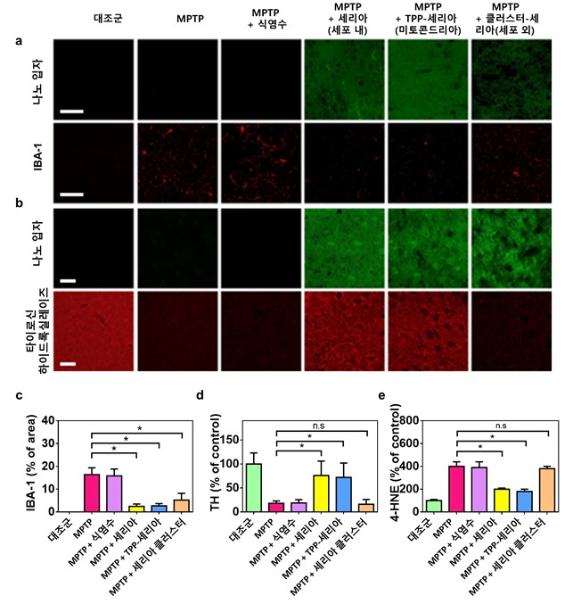
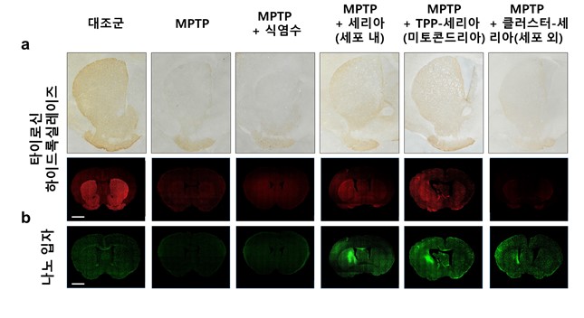
The experiment that the team conducted on mice have demonstrated the effectiveness of the CeO2 nanoparticles. When either the first or the second type of the CeO2 nanoparticles was injected into the striatum of a mouse with Parkinson’s disease, the level of TH (tyrosine hydroxylase), an enzyme involving in the production of neurotransmitter dopamine, could be maintained at comparable levels of normal mice. On the other hand, it was confirmed that the cluster nanoparticles could not protect the TH and only the neuroinflammation had been alleviated.
Professor Hyeon comments, “This finding is the first case to provide localized treatment to Parkinson’s disease and we look forward for its application in the medical field.”
The research findings have been published online on an international journal in the field of chemistry, Angewandte Chemie International Edition (a German applied chemistry journal), on June 22nd. https://onlinelibrary.wiley.com/doi/10.1002/anie.201805052
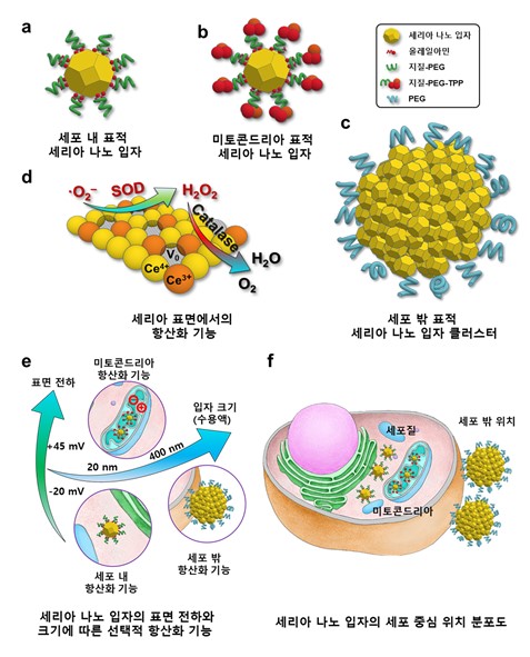
▲ Figure 1. CeO2 Nanoparticles Targeting the Outside, Inside, and Mitochondria of Cell
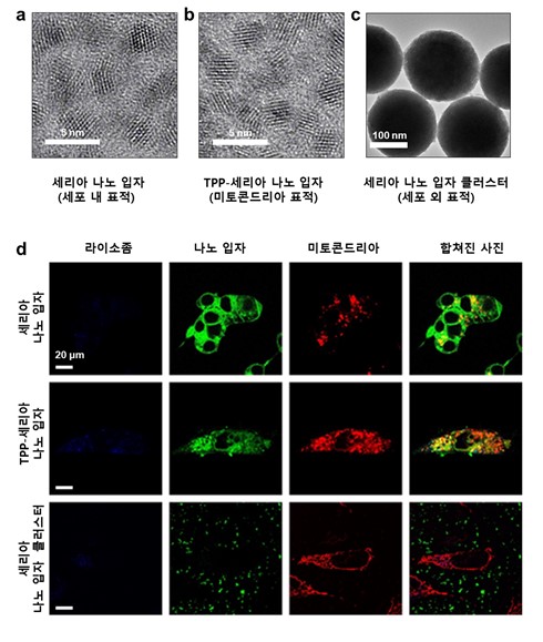
▲ Figure 2. Images of the CeO2 Nanoparticles Obtained from Electron Microscope and Confocal Microscope
▲ Figure 3. Images of Corpus Striata of Mice with MPTP-induced Parkinson’s disease Obtained from Confocal Microscope
▲ Figure 4. Comparison between the TH Levels of Mice With and Without the CeO2 nanoparticle treatment